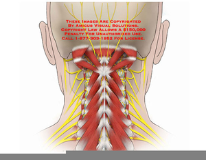
Anatomy Of Back Of Neck : Laminated Cervical Spine Anatomy Poster | Sweat Institute : Some important structures contained in or passing through the neck include the seven cervical vertebrae and enclosed spinal cord, the jugular veins and carotid arteries, part of the esophagus, the larynx.
byAdmin-
0
Anatomy Of Back Of Neck : Laminated Cervical Spine Anatomy Poster | Sweat Institute : Some important structures contained in or passing through the neck include the seven cervical vertebrae and enclosed spinal cord, the jugular veins and carotid arteries, part of the esophagus, the larynx.. Some important structures contained in or passing through the neck include the seven cervical vertebrae and enclosed spinal cord, the jugular veins and carotid arteries, part of the esophagus, the larynx. Anatomy of the nervous system. The splenius muscles originate at the midline and run laterally and superiorly to their insertions. Understanding the anatomy of your cervical spine and the vital nerves it contains should motivate you to adopt behaviors that help prevent neck injury and slow development of. Clinically, surface anatomy is used to split the neck into anterior and posterior triangles which provide clues as to the location of specific structures.
Neck muscles help support the cervical spine and contribute to movements of the head, neck, upper back, and posterior longitudinal ligament (pll). Click now to study the muscles, glands and organs of the neck at kenhub! 12 photos of the anatomy of the back of the neck. « back show on map ». An mri of the face and neck was performed on a healthy patient.
Best Head And Neck Anatomy Stock Photos, Pictures ... from media.istockphoto.com Anatomy of the infrahyoid neck. Anatomy of the hand overview. Some important structures contained in or passing through the neck include the seven cervical vertebrae and enclosed spinal cord, the jugular veins and carotid arteries, part of the esophagus, the larynx. Neck, in land vertebrates, the portion of the body joining the head to the shoulders and chest. In the neck, the platysma when contracted throws the skin into oblique ridges parallel with the fasciculi of the muscle. The pll starts at c2 and goes down the back of the vertebral bodies and intervertebral discs. The structure is, of course, an important part of the conversation. A fractured neck of femur (nof) is a common orthopaedic presentation.
Left (by josh reed), right row 3 left, middle, right row 4 row 5:
Anatomy of male back and neck pain in blue stock. The physicians originally studying human anatomy thought the skull looked like an helmet. In radiology, the 'head and neck' refers to all the anatomical structures in this region excluding the central nervous system, that is, the brain and spinal co. 12 photos of the anatomy of the back of the neck. This entry was posted in anatomy by admin. Jugularis they unite with small veins from the deep muscles at the upper part of the back of the neck, and form a vessel which enters the foramen in the transverse. Traditionally the anatomy of the infrahyoid neck has been subdivided into a group of surgical triangles whose borders are readily palpable bones and. Join our newsletter and receive our free ebook: Over 65000 hip fractures each year are recorded occur in the uk alone and they are becoming in this article, we will look at the classification, anatomy, clinical and radiological features, and management of neck of femur fractures. Want to learn more about it? Anatomy of the back top image row 2: The structure is, of course, an important part of the conversation. This article describes the anatomy of the head and neck of the human body, including the brain, bones, muscles, blood vessels, nerves, glands, nose, mouth, teeth, tongue, and throat.
The anterior jugular vein (v. The splenius muscles originate at the midline and run laterally and superiorly to their insertions. If you'd like to support us and get something great in return, check out our osce checklist booklet containing over 120 osce checklists head & neck anatomy. Whether it's to pass that big test, qualify for that big promotion or even master that cooking technique; The splenius muscles originate at the midline and run laterally and superiorly to their insertions.
Posterior Cervical Anatomy | Free Images at Clker.com ... from www.clker.com A fractured neck of femur (nof) is a common orthopaedic presentation. Despite being a relatively small region, it contains a range of important anatomical features. Your neck is like no other part of the vertebral spinal column and enables your head and neck a wide range of motion. If you'd like to support us and get something great in return, check out our osce checklist booklet containing over 120 osce checklists head & neck anatomy. The splenius muscles originate at the midline and run laterally and superiorly to their insertions. Left (by josh reed), right row 3 left, middle, right row 4 row 5: Over 65000 hip fractures each year are recorded occur in the uk alone and they are becoming in this article, we will look at the classification, anatomy, clinical and radiological features, and management of neck of femur fractures. The infrahyoid neck is the region of the neck extending from the hyoid bone to the thoracic inlet.
Jugularis they unite with small veins from the deep muscles at the upper part of the back of the neck, and form a vessel which enters the foramen in the transverse.
The infrahyoid neck is the region of the neck extending from the hyoid bone to the thoracic inlet. Some important structures contained in or passing through the neck include the seven cervical vertebrae and enclosed spinal cord, the jugular veins and carotid arteries, part of the esophagus, the larynx. Dummies helps everyone be more knowledgeable and confident in applying what they know. Clinically, surface anatomy is used to split the neck into anterior and posterior triangles which provide clues as to the location of specific structures. From a topographical standpoint, there are six major muscle groups in the neck. Jugularis they unite with small veins from the deep muscles at the upper part of the back of the neck, and form a vessel which enters the foramen in the transverse. Dummies has always stood for taking on complex concepts and making them easy to understand. The cervical spine supports the weight and movement of your head and protects the nerves exiting your brain. Our neck is where we find the seven cervical vertebrae, with c7 (the seventh cervical vertebra) meeting t1 (the first thoracic vertebra) at the base of the neck. An mri of the face and neck was performed on a healthy patient. Learn about the various causes of back pain, including different kinds of arthritis. Neck muscles help support the cervical spine and contribute to movements of the head, neck, upper back, and posterior longitudinal ligament (pll). Your neck is like no other part of the vertebral spinal column and enables your head and neck a wide range of motion.
Dummies has always stood for taking on complex concepts and making them easy to understand. Spine anatomy back neck pain tyler tx. Left (by josh reed), right row 3 left, middle, right row 4 row 5: Clinically, surface anatomy is used to split the neck into anterior and posterior triangles which provide clues as to the location of specific structures. Anatomy of the nervous system.
Mouth and neck anatomy, 1866 illustration - Stock Image ... from media.sciencephoto.com Dummies helps everyone be more knowledgeable and confident in applying what they know. Massage therapy for upper back pain. The splenius muscles originate at the midline and run laterally and superiorly to their insertions. Click now to study the muscles, glands and organs of the neck at kenhub! The physicians originally studying human anatomy thought the skull looked like an helmet. Anatomy of the infrahyoid neck. This article describes the anatomy of the head and neck of the human body, including the brain, bones, muscles, blood vessels, nerves, glands, nose, mouth, teeth, tongue, and throat. Want to learn more about it?
From the sides and the back of the neck, the splenius capitis inserts onto the head region, and the splenius cervicis extends onto the cervical region.
From a topographical standpoint, there are six major muscle groups in the neck. The head rests on the top part of the vertebral column, with the skull joining at c1. Massage therapy for upper back pain. 12 photos of the anatomy of the back of the neck. Left (by josh reed), right row 3 left, middle, right row 4 row 5: Clinically, surface anatomy is used to split the neck into anterior and posterior triangles which provide clues as to the location of specific structures. Whether it's to pass that big test, qualify for that big promotion or even master that cooking technique; Understanding the anatomy of your cervical spine and the vital nerves it contains should motivate you to adopt behaviors that help prevent neck injury and slow development of. Dummies has always stood for taking on complex concepts and making them easy to understand. Resists back hyperextension, c1 to sacrum resists hyperflexion of the back, helps prevent herniation, c2… Traditionally the anatomy of the infrahyoid neck has been subdivided into a group of surgical triangles whose borders are readily palpable bones and. Head and neck anatomy is important when considering pathology affecting the same area. We've largely focused on the physical aspect of our spinal anatomy in this series.